Examples
What can you do with « PET quantif »?
1. Image denoising for improved quantification
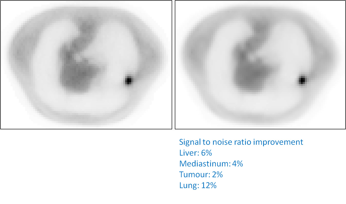
On the left, an axial slice of a PET image with a small tumor in the lungs. On the right, the image filtered with the optimized denoising approach combining wavelets and curvelets tools. Notice how the homogeneous areas (lungs, mediastinum) have been filtered and are less noisy, while the tumor and the organs contours have not been significantly altered, contrary to what is usually obtained using post-reconstruction Gaussian filtering.
2. Image deconvolution for Partial Volume Effects correction, both visual and quantitative correction
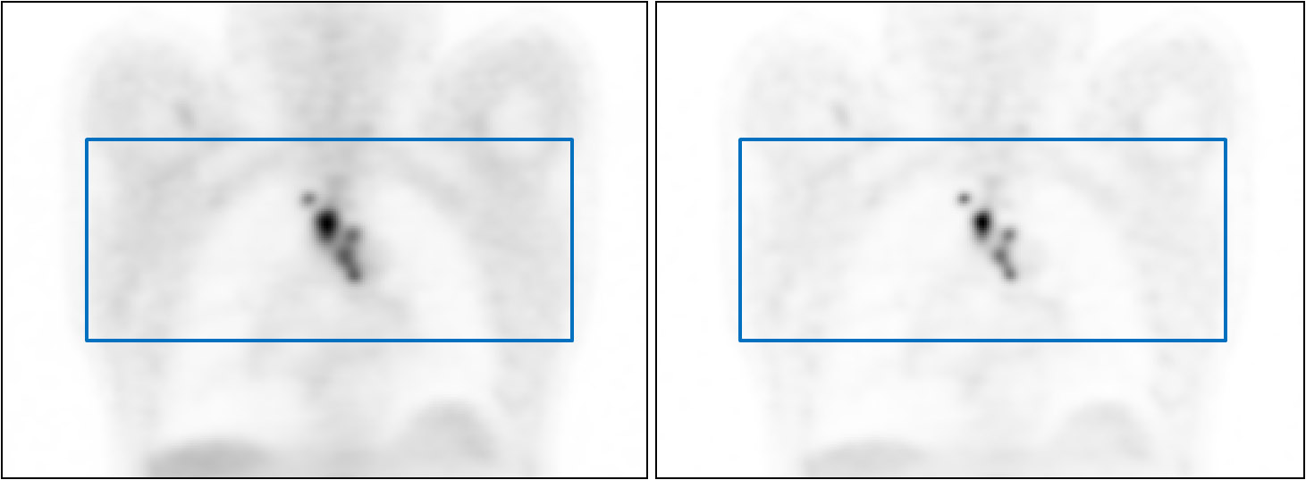
On the left, a coronal slice of a 18F-FDG PET image of an esophageal lesion. On the right, the corresponding deconvolved image using iterative deconvolution combined with improved wavelet based denoising within the iterative process. Below, the quantitative improvement obtained expressed as tumor to background ratios and profiles:
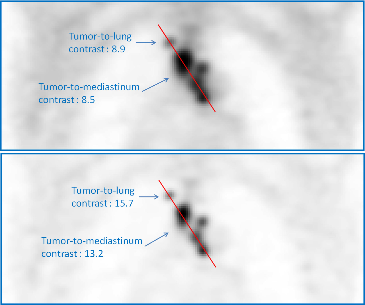
Notice how the various uptakes are much more differentiated in the deconvolved image corrected for Partial Volume Effects. Overall contrast is also significantly improved without significant addition of noise (which is often the case with standard iterative deconvolution).
3. Metabolically active tumor volume (MATV) automatic delineation, either on original or filtered (denoising, deconvolution) images for full assessment of tumor.
MATV can be automatically delineated in 3D on PET images using the Fuzzy Locally Adaptive Bayesian (FLAB) method, as illustred below on both the original and the deconvolved PET images used above.
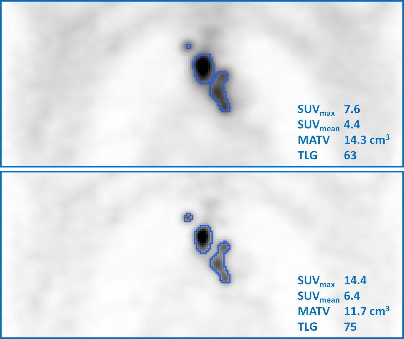
Notice how the delineation is both pertinent with respect to the content of the image. Parameters associated to the delineated MATV (SUV measurements or total lesion glycolysis) can be automatically provided for further analysis.
FLAB can also be used for radiotherapy planning, for which its ability to delineate sub-volumes can be exploited for dose boosting or dose painting scenarios as shown below:
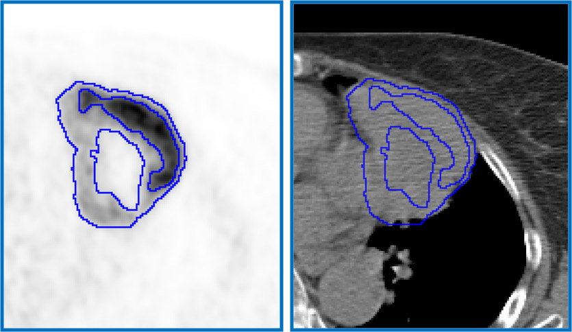
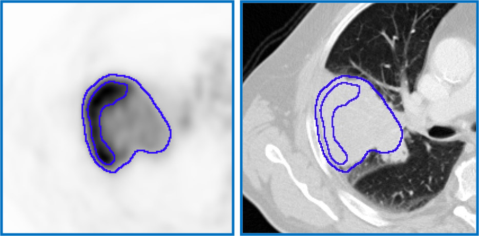
Above, two examples of large NSCLC tumors exhibiting heterogeneous PET tracer uptake. FLAB is able to automatically provide external contours of the MATV as well as sub-volumes contours within the MATV.